Ever wondered what Year 12 Biologists get up to?
This is what they’ve been doing this week:
1. Investigating the Transpiration-Cohesion Hypothesis
Cohesion hypothesis is a generally accepted explanation of the rise of sap in plants by means of intermolecular attractions. Calculation and experiment indicate that the forces of cohesion between water molecules and the forces of adhesion between water molecules and the walls of the xylem vessel cells are sufficient to confer on thin columns of water a tensile strength of at least 30 atmospheres (440 pounds per square inch). This is high enough to permit a thin column of water to be lifted to the top of any tree without breaking the column.
The cohesion of water explains only maintenance of the sap column; the explanation for the upward movement of the water is accounted for by a mechanism, called transpiration pull, that involves the evaporation of water from leaves. Thus, the explanation for the upward movement of sap in trees is also called the transpiration-cohesion hypothesis.
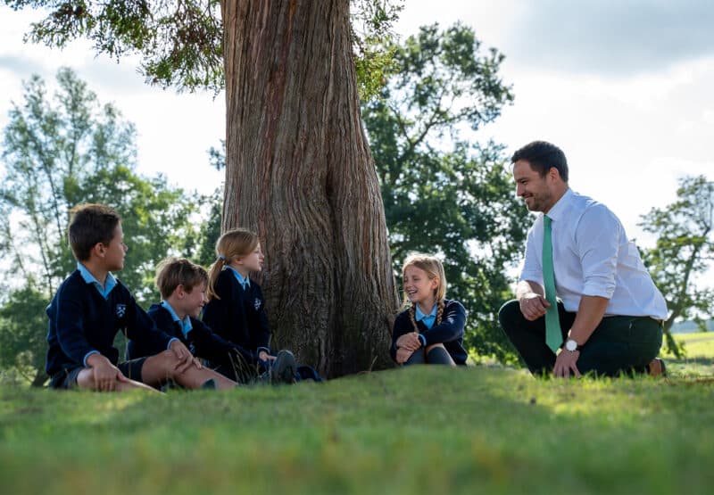
Year 12 Biologists in class, yesterday, modelled the transpiration-cohesion hypothesis by using straws, to represent xylem tubes, and themselves to represent transpiration! They were not as successful as most plants are!
![]()
Laetitia, Fred and Charlotte testing the cohesion – transpiration theory
2. Dissection and microscopy of plant vascular tissue
Year 12 Biologists have also been examining the structure of the vascular bundle (xylem and phloem) in plants. In one experiment students used celery to produce and observe a stained specimen under the microscope. The use of the stain toluidine blue provided a colour difference between lignified and non-lignified cell walls; clearly highlighting specialised cells. Whilst a reasonably simple experiment, it is hard to produce a really thin specimen which clearly shows all the features, but Gaby has produced a really beautiful, almost textbook level photomicrograph of the vascular bundle in celery at x 400 magnification.

Dissection and microscopy of plant vascular tissue by Gaby, Y12, Biology
3. Xylem Tubes
Charlotte, Year 12 Biologist, produced this lovely photomicrograph in our lesson yesterday, showing xylem tubes in rhubarb. The arrangement of the lignin, in the spiral and annular arrangements, in the xylem tubes can be clearly seen in this photomicrograph at x 400 magnification.

Charlotte’s impressive photomicrograph